What happens when an essential nutrient overshoots its welcome—like a well-aimed firework that turns into a bonfire? Vitamin D toxicity is exactly that kind of clinical “plot twist.” It can arrive silently, then suddenly announce itself with hypercalcemia, dehydration, and an assortment of symptoms that feel loosely connected—until you understand the chemistry behind them. The management strategy is both rigorous and oddly elegant: you treat the elevated calcium, you respect the physiology, and you nudge the body back toward equilibrium. But before you reach for a treatment checklist, ask yourself a playful question: Could your patient’s “just one more supplement” be the villain?
Recognizing the toxicity: when vitamin D turns mischievous
Vitamin D toxicity typically reflects prolonged or excessive intake of vitamin D or its metabolites, leading to persistently elevated serum calcium. Hypercalcemia, in turn, can create a cascade—vomiting, constipation, confusion, polydipsia, polyuria, and fatigue. Some patients appear oddly sluggish, like their nervous system is running on low voltage. Others present with musculoskeletal discomfort or neurocognitive changes, which can be mistaken for unrelated illness.
Clinically, the pattern matters. Consider the timeline: a supplement history, fortified products, or accidental double-dosing can set the stage. Laboratory evaluation often reveals high calcium, suppressed PTH, elevated 25-hydroxyvitamin D (sometimes with additional metabolite details), and variable renal involvement. Kidney injury is a common subplot—hypercalcemia can precipitate nephrocalcinosis and compromise filtration.
Potential challenge: symptoms can mimic infection, gastrointestinal disorders, or dehydration from other causes. The playful part ends when you realize dehydration is not merely a side effect—it can be the accelerator pedal for toxicity. In other words, the body can become trapped in a cycle of elevated calcium and worsening volume status.
First principles of management: stop the supply, stabilize the terrain
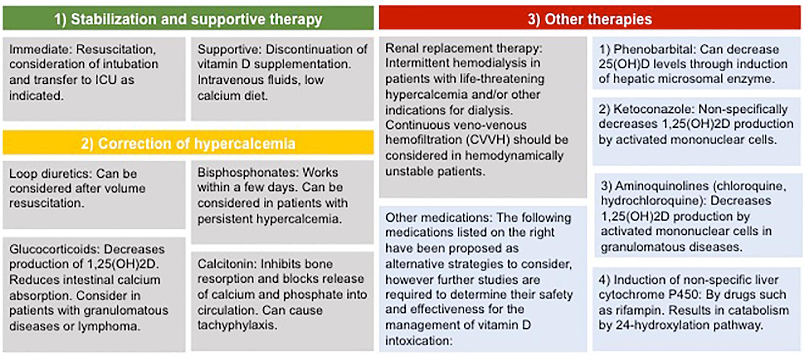
Management begins with immediate cessation of vitamin D sources, including supplements and high-dose therapeutic prescriptions. If the exposure was accidental, the lesson is not only clinical; it’s logistical. Ensure med lists are accurate, and review all over-the-counter products—many preparations contain vitamin D in surprising quantities.
Next comes stabilization. Hypercalcemia is a physiologic stressor that can affect the heart and brain. If there are severe symptoms—altered mental status, marked dehydration, or significantly elevated calcium—treat as urgent. Even when the patient looks “okay,” the calcium level can still be quietly rewriting organ physiology.
As you move through the plan, you’ll notice a theme: the body needs volume, then it needs a mechanism to lower calcium, and finally it needs monitoring to prevent rebound.
IV fluids: the unsung hero that resets the calcium equation
Intravenous isotonic fluids are often the first definitive maneuver. They restore intravascular volume, correct dehydration, and enhance renal excretion of calcium. Think of IV fluids as opening a clogged irrigation channel—suddenly the kidneys can clear the calcium load. This is especially important when nephrogenic mechanisms are compromised by volume contraction.
Typically, clinicians use isotonic saline and adjust the rate to the patient’s circulatory status, renal function, and electrolyte profile. The goal is not “more fluid,” but the right fluid in the right amount at the right time.
Electrolytes must be monitored. Hypernatremia, hypokalemia, or evolving renal impairment can complicate treatment. Potassium balance deserves respect, because hypokalemia may worsen urinary concentrating defects and influence overall metabolic stability. In severe cases, fluids may be continued until calcium trends downward and symptoms improve.
Diuretics and urine strategy: why pushing urine is not the first move
Sometimes after adequate hydration, clinicians consider loop diuretics to promote calciuresis. But timing matters. Using diuretics before restoring volume can worsen dehydration and intensify hypercalcemia. It’s like turning up the pressure in a leaky system—initially it may look productive, but it can accelerate harm.
Loop diuretics can increase urinary calcium excretion once the patient is properly rehydrated. The strategy depends on urine output, hemodynamics, and kidney function. Ongoing monitoring of electrolytes and hydration status remains essential.
In practice, the diuretic phase feels like a second act: first you build the foundation, then you sweep calcium out through renal pathways.
Steroids: the molecular dampers for certain causes
Here’s where the story gets fascinating. Corticosteroids can help in cases of vitamin D toxicity when the physiology involves increased vitamin D activity beyond simple intake—such as granulomatous diseases (e.g., sarcoidosis) or certain lymphoproliferative conditions where extra renal production of active vitamin D metabolites occurs. Steroids reduce intestinal absorption of calcium and may suppress production of active vitamin D metabolites.
Clinically, steroids can become a pivot point when standard hydration alone is insufficient or when the underlying mechanism suggests immune-mediated overproduction of vitamin D activity. Dose selection varies with severity, response, and comorbidities.
The challenge is that steroid therapy is not a universal key. It’s an intentionally targeted tool, best matched to the presumed pathophysiology. When used appropriately, it can quiet the biochemical noise driving hypercalcemia.

Bisphosphonates: when calcium needs a stronger brake
If hypercalcemia is severe, persistent, or symptomatic despite fluids (and possibly steroids), clinicians often consider bisphosphonates. These medications inhibit osteoclast-mediated bone resorption, thereby reducing the release of calcium into circulation. It’s a kind of skeletal “pause button,” limiting the body’s contribution to circulating calcium elevation.
Bisphosphonates are especially useful when the hypercalcemia is refractory or when rapid control is required to mitigate complications. Renal function must guide selection and dosing, as these agents depend on kidney clearance and can pose risks in patients with impaired filtration.
Response is usually not instantaneous, but it can be meaningful—calcium frequently begins to fall over the next day(s). Monitoring continues because the body’s calcium regulation can be nonlinear, and treatment effects may lag behind symptoms.
Calcitonin and refractory scenarios: temporary relief with tactical purpose
Calcitonin may be used for short-term management, particularly when there is an urgent need to lower calcium while waiting for other therapies to take full effect. It can offer early reduction in serum calcium, acting as a bridge therapy.
Its effect may wane over time due to tachyphylaxis, so clinicians typically treat it as a tactical interlude rather than the whole resolution. Still, in a crisis—when the patient is symptomatic—bridging can be the difference between stabilization and deterioration.
Dialysis and advanced rescue: the “last resort” that still matters
In extreme cases—especially with profound renal failure or life-threatening hypercalcemia—hemodialysis can be considered. It can remove calcium and correct metabolic disturbances rapidly. While it sounds dramatic, dialysis is a rational option when the kidneys cannot participate in detoxification.
This pathway requires careful coordination with nephrology and continuous monitoring for complications. It’s not everyday medicine, but it can be life-saving for the right patient at the right moment.
Correcting the electrolytes: potassium, phosphate, magnesium, and the hidden details
Hypercalcemia treatment is never just about calcium. As calcium levels change, other electrolytes can follow their own shifting patterns. Potassium, phosphate, and magnesium require attention. Hypokalemia can worsen weakness and complicate arrhythmia risk. Magnesium abnormalities can influence calcium handling and neuromuscular stability.
Electrolyte correction should be integrated into the management plan, not treated as an afterthought. The body is a tightly coupled system—adjust one variable and others react.
Monitoring and preventing rebound: the aftercare chapter
Vitamin D is fat-soluble, which means that once it’s stored in adipose tissue, it may be released gradually. This can lead to delayed or recurrent hypercalcemia even after initial improvement. Monitoring should therefore continue beyond the early stabilization window.
Serial calcium measurements guide the pace of treatment tapering. Symptoms should be reassessed frequently. Kidney function and urine output should remain under surveillance, particularly if there was early injury.
Finally, long-term prevention is the real moral of the story. Review supplementation habits, educate about dosing, and ensure that vitamin D intake aligns with clinical indications. The most effective “therapy” often lies in preventing the initial overshoot.
Putting it together: a cohesive algorithm for real patients
So how does the management sequence typically unfold? First, stop the vitamin D and assess severity. Then restore volume with isotonic IV fluids. If needed, consider diuretics after rehydration, add steroids when the mechanism supports immune-mediated metabolite activity, and escalate to bisphosphonates for persistent or severe cases. Calcitonin can bridge acute crises, and dialysis becomes a rescue tool when the kidneys cannot safely clear calcium.
Through all of it, monitoring is the quiet conductor—electrolytes, renal function, ECG risk, and symptom evolution are the score. The playful question returns: Could “just vitamin D” be a disguised emergency? In the right context, yes. But with a structured approach—fluids, targeted anti-inflammatory therapy, calcium-lowering agents, and vigilant follow-up—the chaos can be converted into control.