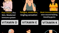
Tooth development is a finely tuned orchestration of minerals, proteins, and cellular signaling. When something nudges that process off course, the results can be frustrating: delayed eruption, enamel that feels thinner, or a surface that seems more vulnerable than expected. One question often rises in conversations about oral health is whether low vitamin D can be a culprit. The answer is nuanced. Vitamin D doesn’t “directly” stain enamel overnight, but it can influence the biochemical environment that makes enamel possible in the first place—and that influence can echo into childhood and beyond.
Vitamin D: The Mineral “Gatekeeper” Behind Enamel Formation
Vitamin D is best known for regulating calcium and phosphate—two minerals that are not optional for tooth mineralization. Enamel, the hardest substance in the human body, is essentially a mineral-rich tissue built through crystal growth and layering. For that to happen reliably, your body must absorb enough calcium and phosphate from the gut and deliver them to developing teeth.
When vitamin D is insufficient, calcium absorption can drop. That can reduce the raw materials available for enamel formation. Even if the body tries to compensate, the developmental timeline may not tolerate repeated shortages. Think of it as trying to build a masonry wall when mortar shipments arrive late: the structure can still rise, but the quality and resilience may vary.
Enamel problems tied to mineral imbalance often reveal themselves through altered opacity, hypomineralization, or structural weaknesses that may increase susceptibility to caries. The timing matters too—developmental stages have different vulnerabilities.
Can Low Vitamin D Cause Delayed Tooth Eruption?
Tooth eruption is a dynamic process, not merely a countdown from childhood. It requires coordinated bone remodeling, periodontal ligament activity, and adequate mineral supply. Since vitamin D contributes to calcium homeostasis and bone metabolism, a deficiency could plausibly influence eruption timing.
Some individuals with low vitamin D experience broader growth-related concerns: delayed skeletal development, altered bone density, or generalized fatigue. In that context, tooth eruption can also shift, particularly when deficiency is prolonged during childhood.
However, delayed eruption has many causes. Genetics, nutritional patterns, chronic illness, endocrine disorders, and congenital dental anomalies all compete for attention. Vitamin D insufficiency may be a contributing factor rather than a solitary explanation.
In practical terms, if a child’s teeth appear late compared with expected eruption ranges, it is wise to consider a broader evaluation: diet quality, overall growth, iron and calcium status, and medical history. A clinician can decide whether vitamin D testing is warranted.
Enamel Problems: How Deficiency May Manifest in Tooth Quality
Enamel forms through progressive mineral deposition. If calcium and phosphate availability is insufficient during the mineralization window, enamel may become hypomineralized—meaning the tissue contains less mineral density and exhibits altered texture and translucency.
Enamel defects can appear as:
• Reduced mineral density that makes enamel more prone to surface wear.
• Opacity changes, where spots look chalky or dull rather than uniformly glossy.
• Increased sensitivity, especially after brushing or temperature changes.
• Higher cavity risk due to compromised structural defenses.
Not all enamel issues are vitamin D–driven. Other possibilities include fluoride exposure variability, premature enamel hypoplasia from early illness, genetic enamel formation defects, and medications that affect mineral balance. Still, vitamin D deficiency can create a permissive environment for these vulnerabilities to become more noticeable.
Different Types of Enamel Issues Readers Might Hear About
Dental discussions often use overlapping terms. Understanding them helps readers interpret symptoms without panic.
Enamel Hypoplasia refers to enamel that formed with fewer minerals or incomplete thickness. It may show pits, grooves, or irregular surface contours. The appearance can look “underdeveloped,” as if enamel didn’t lay down adequately during early formation.
Enamel Hypomineralization describes enamel that formed with normal structure but reduced mineral content. It can look chalky or more opaque and may lead to brittle enamel that fractures more easily.
Developmental Dental Defects is a broader category. It includes defects influenced by systemic conditions during tooth development, including nutritional shortfalls and metabolic disruptions. Vitamin D is one variable within this category.
When people say “enamel problems,” they may be referring to sensitivity, visible discolorations, or frequent cavities. Each scenario can have different underlying biology.
Symptoms and Clues: When Low Vitamin D Becomes Relevant to Oral Health
Vitamin D deficiency doesn’t announce itself solely through teeth. It tends to show up in the body first. Common systemic clues include bone or muscle aches, frequent fatigue, and a general vulnerability to infections. In children, it can also correlate with growth concerns.
Oral clues may include increased frequency of cavities, enamel that seems to wear down faster, and sensitivity that appears earlier than expected. Some individuals notice discolorations—patches that look lighter, chalky, or uneven.
That said, the mouth is not a medical diagnostic screen. Many enamel changes also stem from factors like early childhood illness, orthodontic trauma, plaque retention, or enamel maturation differences after eruption.
Still, if oral changes align with risk factors for vitamin D deficiency—such as limited sun exposure, darker skin pigmentation in high-latitude regions, strict dietary restrictions, or breastfeeding without supplementation—the connection becomes more plausible.
Who Is at Higher Risk of Low Vitamin D?
Low vitamin D is more common than many people expect. Risk increases when sun exposure is limited, when diets lack vitamin D–rich foods, or when absorption is impaired. People living indoors, working night shifts, or using consistent high-SPF sun protection may also be at risk.
Certain medical conditions—especially those affecting the gastrointestinal tract—can reduce absorption of fat-soluble vitamins. Similarly, some medications may alter vitamin D metabolism over time.
In children, inadequate vitamin D can be especially consequential because teeth and bones are actively developing. The gap between deficiency and dental outcomes can be long enough that parents attribute changes to “normal development,” even when a metabolic factor was present.
Testing and Clinical Evaluation: What Readers Can Expect
Clinicians usually confirm vitamin D status with a blood test, often measuring 25-hydroxyvitamin D. If the result is low, medical history and dietary patterns are reviewed. Calcium, phosphate, and sometimes parathyroid hormone may also be considered to understand how the body is responding.
For dental concerns such as suspected enamel defects, a dental exam can reveal patterns. A dentist may assess tooth surfaces, evaluate whether defects are consistent across multiple teeth, and review eruption timing. Sometimes imaging or enamel evaluation tools are used, depending on the case.
Importantly, evaluation should not stop at vitamin D alone. If enamel issues appear severe or localized, other causes may be more likely, including genetic enamel disorders or early systemic illness.
Treatment and Prevention: Can Vitamin D Improve Existing Enamel?
Vitamin D supplementation primarily supports mineral metabolism during development. Once enamel is fully formed, its ability to “heal” is limited. Enamel does not regenerate like skin. However, improving vitamin D status can help protect future tooth development and supports overall oral health.
For existing enamel weakness, prevention becomes the main strategy. Fluoride exposure, careful brushing technique, dietary counseling to reduce sugar frequency, and dental sealants or restorative interventions can reduce the risk of cavities and fractures.
If a deficiency is corrected early, the prospects for healthier enamel formation in developing teeth improve. The strongest benefit is often seen when supplementation aligns with the developmental window of tooth mineralization.
Oral Care Steps That Complement Nutritional Support
When readers connect vitamin D with tooth delay or enamel problems, it is helpful to pair medical correction with high-impact dental habits.
Consider:
• Fluoride toothpaste to strengthen enamel against acid challenges.
• Reduced sugar “spikes” through consistent meal timing rather than frequent snacking.
• Gentle brushing with appropriate technique to protect porous enamel surfaces.
• Dental checkups focused on early caries detection and enamel vulnerability assessment.
• Sealants for at-risk molars, depending on the dental evaluation.
These actions don’t replace supplementation when deficiency exists. They simply offer immediate mechanical and chemical protection while the body’s mineral systems recalibrate.
When to Seek Help: Red Flags and Timing
Seek professional guidance if tooth eruption is significantly delayed, if enamel appears chalky or frequently chips, or if cavities develop unusually fast despite good brushing habits. For children, it is especially important not to wait too long—early evaluation can clarify whether a systemic factor like vitamin D deficiency is involved.
Also consider medical consultation if vitamin D deficiency risk is high: limited sun exposure, dietary constraints, or symptoms suggestive of broader deficiency. Teeth can be an early “story chapter,” but the full narrative belongs to the body.
Conclusion: A Plausible Link, Best Understood as Part of a Bigger System
Low vitamin D can contribute to tooth development issues by influencing calcium and phosphate availability—factors that shape enamel mineralization and support normal growth processes. This means vitamin D deficiency may be associated with delayed eruption and enamel defects, especially when the deficiency persists during the developmental years.
Yet enamel problems are rarely one-cause mysteries. They can arise from genetics, early illnesses, nutritional patterns, and other metabolic influences. The most effective path is a coordinated approach: evaluate vitamin D status when risk factors or symptoms align, and pair that with targeted dental prevention to protect vulnerable enamel.